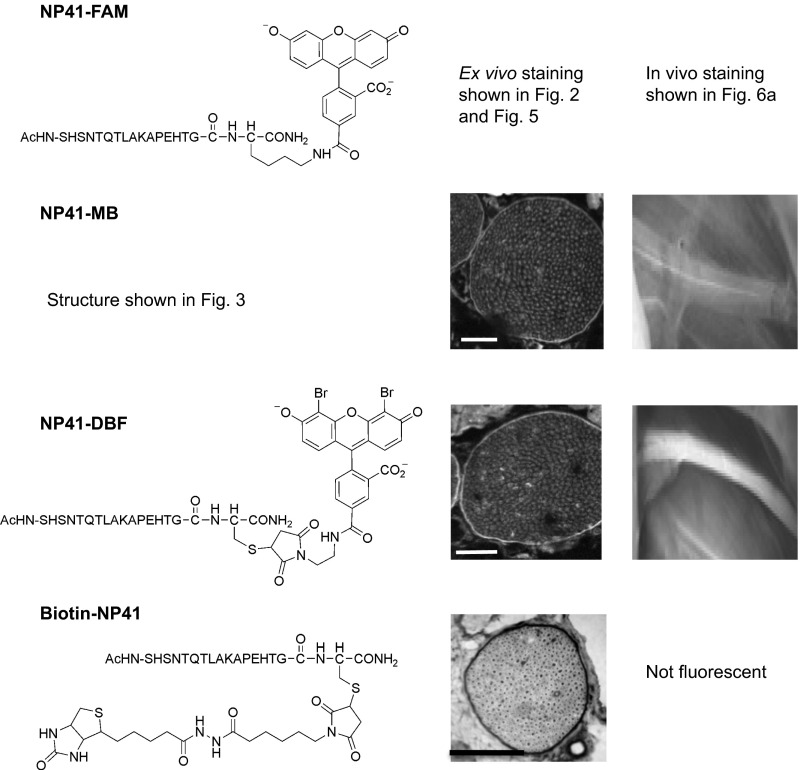
Fig. S3.
NP41 conjugated to various SOGs and biotin shows consistent perineurial localization, whereas in vivo nerve-to-muscle contrast differs between conjugates. (Left) NP41 conjugates and structures. (Middle) NP41–MB and NP41–DBF treated on nerve sections and imaged by fluorescence show perineurial staining (highlighted areas). NP41–biotin treated on tissue sections and stained by streptavidin–HRP and DAB shows strong perineurial signal (darkened areas). (Scale bar, 500 mm.) Gain levels are set individually. (Scale bar, 50 mm.) (Right) NP41 dyes, injected systemically into mice and imaged 2–4 h after, show different sciatic nerve contrast in vivo. NP41–MB showed low nerve fluorescence in vivo that was difficult to distinguish from the surrounding muscle. NP41–DBF showed higher nerve fluorescence and contrast to muscle (n = 2).

