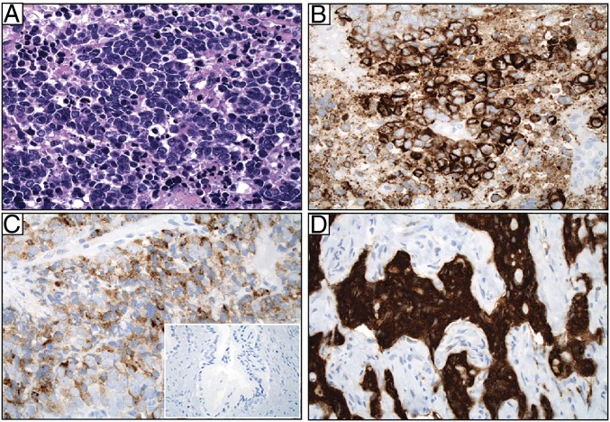
Fig. 2.
GRP78 protein is expressed both in the cytoplasm and at the cell surface in AVPC specimens from patients. (A) Pathological evaluation of a representative specimen with high-grade cytology, small cell features, nuclear molding, and necrosis (H&E). (B) The same tumor stained for GRP78, moderate-to-strong (2–3+) intensity in tumor cells. (C) GRP78 staining of a representative case showing weak (1+) but diffuse positivity. (D) GRP78 staining of a representative case showing strong (3+) intensity. (Inset) Staining of normal prostate gland. (Magnification: A–D, 40×; Inset, 5×.)

