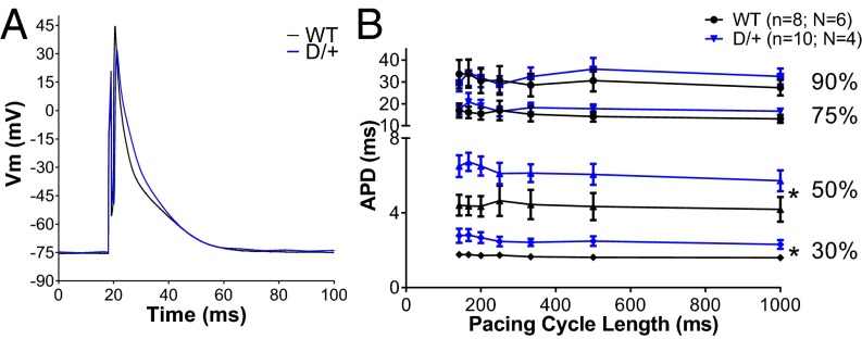
Fig. 2.
Scn8aN1768D/+ myocytes have altered AP morphology. (A) Representative traces from WT (black) and Scn8aN1768D/+ (red) ventricular myocytes. (B) The early stages of repolarization (APD30 and APD50) are significantly prolonged in Scn8aN1768D/+ myocytes compared with WT (P = 0.01 and P = 00.04, respectively). The total APD, APD90, is not altered (P = 0.41). Scn8aN1768D/+ (D/+): n = 10, N = 4; and Scn8a+/+ (WT): n = 8, N = 6. Vm, membrane potential. *P < 0.05 vs. WT.

