Figure 3.

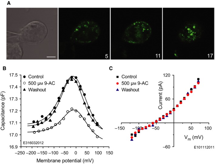
9‐AC reversibly reduces the NLC of prestin‐transfected HEK 293 cells. (A) Prestin‐transfected HEK 293 cell possessing the typical dotted fluorescence in the plasma membrane. Cells were voltage‐clamped using 135 mm CsCl intracellular solution (upper left/DIC image). GFP labelling was recorded with confocal microscopy using an argon laser with excitation wavelength of 488 nm. The emitted light was detected above 505 nm. Numbers in the bottom right corners of the fluorescence images indicate the level of the confocal section in microns. (B) Capacitance of the prestin‐transfected HEK 293 cell in A. Extracellular application of 500 μm 9‐AC reversibly reduces the NLC. Lines indicate fits using Eqn (1) with parameters given in Table 5. (C) I‐V characteristics of a non‐transfected HEK 293 cell, indicating that 9‐AC does not influence voltage‐gated chloride conductances in this cell line. Scale bar, 10 μm.
