
Figure 5.
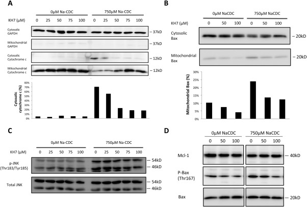
sAC inhibition prevented cytochrome c release and mitochondrial translocation of Bax during BSIA. (A) Cytochrome c release assay. H69 cholangiocytes were treated with increasing concentrations of KH7 in the absence or presence of 750 μM NaCDC for 1 hour in a 5% CO2, 37°C incubator. Cell fractionation was performed as described in Materials and Methods. Lysates of equal volume were subjected to sodium dodecyl sulfate‐polyacrylamide gel electrophoresis and immunoblotted for glyceraldehyde 3‐phosphate dehydrogenase, a cytosolic marker, and cytochrome c. Cytochrome c immunoblots were quantified by ImageJ. Ratios of cytosolic signal to total signal (cytosolic plus mitochondrial) were calculated. (B) Bax translocation assay was performed and quantified as in (A). Percentage of mitochondrial Bax was calculated as mitochondrial signals divided by the sum of cytosolic and mitochondrial signals. (C) H69 cholangiocytes were treated as in (A). Whole‐cell lysates were used for immunoblot of total and phosphorylated JNK. (D) H69 cholangiocytes were treated as in (B). Whole‐cell lysates were used for immunoblot for myeloid cell leukemia 1, phospho‐Bax (Thr167), and total Bax. The figure shows a representative experiment from a series with similar results. Abbreviations: GAPDH, glyceraldehyde 3‐phosphate dehydrogenase; Mcl‐1, myeloid cell leukemia 1.