
Figure 4. Architecture of crRNA-Guided and dsDNA Target-Bound Cascade/I-C Complexes.
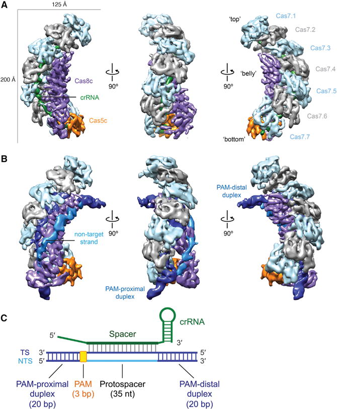
(A and B) Cryo-EM reconstructions of intact apo-Cascade/I-C (crRNA-bound) (A) and “2× duplex” dsDNA target-bound Cascade/I-C (B) at 6.7- and 8.8-Å resolution, respectively. Subunits are segmented and colored as indicated. Details of these structures can be seen in Movies S1 and S2.
(C) Schematic of crRNA and target dsDNA used for cryo-EM analysis. TS, target strand; NTS, non-target strand; PAM, protospacer-adjacent motif.
See also Figures S3 and S4 and Tables S1 and S2.