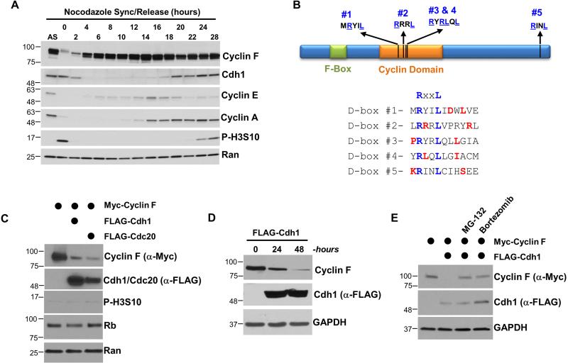
Figure 1. Cdh1 regulates the abundance and stability of Cyclin F.
(A) U2OS cells were synchronized in mitosis with nocodazole, isolated by “shake-off”, and analyzed by immunoblot after release into the cell cycle. (B) Domain structure of human Cyclin F showing the position of its Cyclin homology domain, F-box domain, and five putative APC/C degron motifs. Preferred residues around the putative D-box motifs are shown in red. (C) FLAG-Cdc20 or FLAG-Cdh1 were ectopically expressed in 293T cells in combination with Myc-Cyclin F. Cells were harvested after 24 hours and analyzed by immunoblot. (D) FLAG-Cdh1 was ectopically expressed in 293T cells and reduced the level of endogenous Cyclin F at 24 and 48 hours after transfection. (E) Myc-Cyclin F and FLAG-Cdh1 were ectopically expressed in 293T cells for 24 hours. Cells were treated with the proteasome inhibitors MG-132 (10 μM) or bortezomib (100 nM) four hours prior to harvesting.

