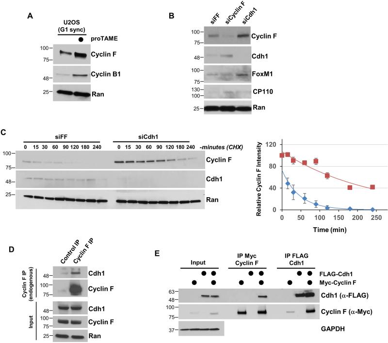
Figure 2. Cyclin F degradation is regulated by the APC/C.
(A) U2OS cells synchronized in G1 phase by mitotic block and release and treated with the APC/C inhibitor proTAME for 90 minutes. (B) Cyclin F or Cdh1 were depleted from T47D cells using siRNA for 48 hours. Negative control siRNA (siFF) targets firefly luciferase. (C) Depletion of Cdh1 using siRNA extended the half-life of Cyclin F. Cells were depleted of Cdh1 by siRNA (same as B), synchronized in G1 by nocodazole block and release, and then treated with cycloheximide (CHX) to analyze Cyclin F stability. On the right is a semi-quantitative analysis of the Cyclin F signal. Negative control (blue diamonds), Cdh1 depletion (maroon squares). Cyclin F signal relative to the Ran loading control is shown on the Y axis (error bars indicate standard deviation of mean). (D) Endogenous Cyclin F was precipitated from 293T whole cell extracts. Cells were treated with MG-132 prior to lysis. (E) Myc-Cyclin F and FLAG-Cdh1 were transfected into 293T cells, and each was separately recovered and analyzed by immunoblot. Cells were treated with MG-132 for four hours prior to lysis.

