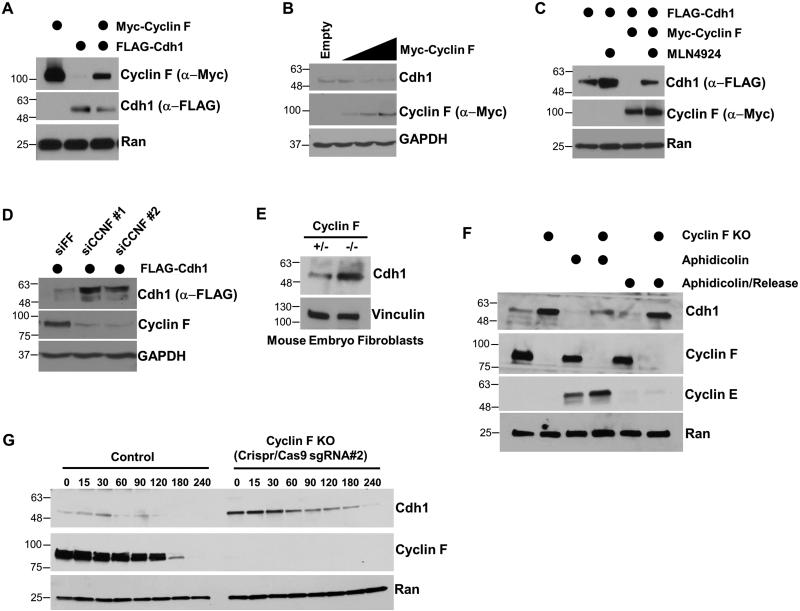
Figure 4. Cdh1 abundance and stability are regulated by Cyclin F.
(A) Ectopic expression of Myc-Cyclin F and FLAG-Cdh1 in 293T cells reduces the level of both proteins. (B) Myc-Cyclin F reduced the abundance of endogenous Cdh1 in a dose dependent manner when expressed in 293T cells. (C) Myc-Cyclin F overexpression reduces the abundance of FLAG-Cdh1 when expressed in 293T cells. The degradation of Cdh1 was rescued by the neddylation inhibitor MLN4924. (D) Depletion of Cyclin F using two independent siRNA reagents increases exogenously expressed FLAG-Cdh1 in 293T cells. (E) Cdh1 levels were analyzed in Cyclin F null (−/−) MEFs and in a corresponding control cell line (+/−). (F) Control and Cyclin F knockout HeLa cells were analyzed either in asynchronous populations (lanes 1 and 2), synchronized in S-phase with aphidicolin (lanes 3 and 4), or 6 hours after aphidicolin release (lanes 5 and 6). During aphidicolin block serum was reduced to mitigate DNA damage. (G) Control and Cyclin F knock-out HeLa cells were treated with cycloheximide and Cdh1 half-life was analyzed.

