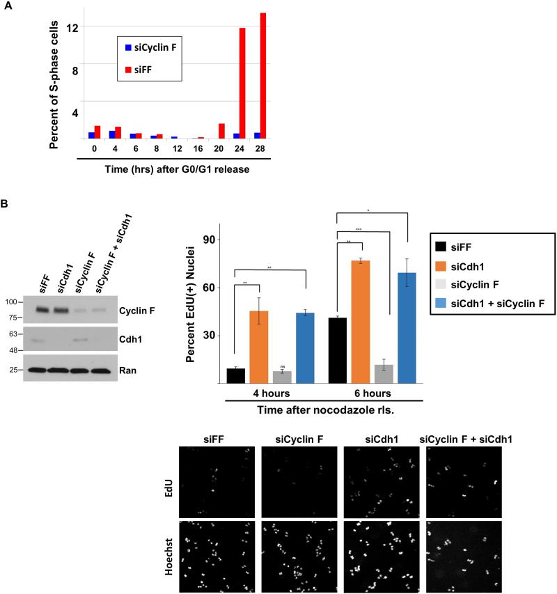
Figure 6. Cyclin F regulation of G1 progression is dependent on Cdh1.
(A) Non-transformed RPE1 cells were treated with siRNA targeting Cyclin F or FF. Cells were synchronized in G0/G1 by serum withdrawal. After refeeding, entry in S-phase was monitored at the designated time points by flow cytometry. (B) U2OS cells were synchronized in mitosis with nocodazole following depletion with siRNA targeting FF, Cdh1, Cyclin F, or Cyclin F and Cdh1 together. After release from nocodazole, cells were pulsed with EdU for 30 minutes, fixed, and analyzed for EdU incorporation. The percent of nuclei that are EdU positive is shown (performed in triplicate, * p≤0.01; ** p≤0.004; *** p≤ 0.0005 ; p values were calculated using un-paired t-test). Immunoblot on left shows knockdown at zero time point (in mitosis). Representative images of EdU positive cells (top) and DNA content (bottom) are shown below.

