Figure 7.
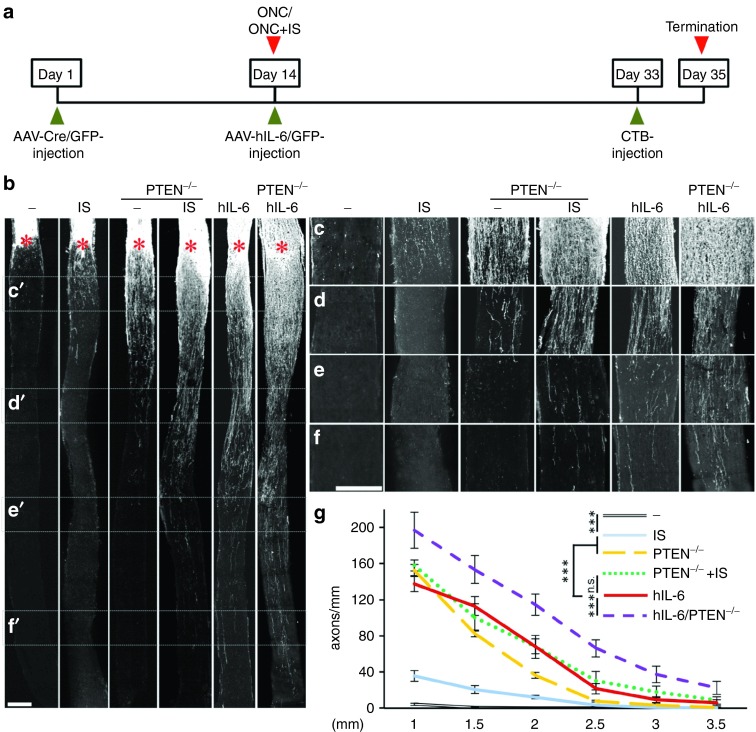
Markedly improved optic nerve regeneration upon hIL-6 expression. (a) Application scheme of the in vivo regeneration studies. Adult floxed PTEN mice were intravitreally injected either with AAV-Cre to induce RGC-specific PTEN deletion (PTEN-/-) or with AAV-GFP (-) as control treatment 14 days prior to optic nerve crush (ONC). ONC was combined either with inflammatory stimulation (IS) or intravitreal injection of either AAV-hIL-6 or AAV-GFP. Fluorescently labeled cholera toxin (CTB) was injected 2 days prior to sacrifice at 3 weeks post ONC. (b) Representative pictures of longitudinal sections of optic nerves isolated from mice treated as depicted in a) with regenerating axons visualized by fluorescent CTB. Asterisks indicate the crush site. Scale bar = 200 µm. (c–f) Magnification of the respective optic nerve sections depicted in b), visualizing regenerating axons at 0.5 (c), 1.5 (d), 2.5 (e), and 3.5 mm (f) from the crush site. Scale bar = 200 µm. At 3.5 mm, axons are only detected in optic nerves of hIL-6, PTEN-/- + IS, and PTEN-/- + hIL-6. (g) Quantification of regenerating axons at 1, 1.5, 2, 2.5, 3, and 3.5 mm beyond the crush site in adult mice treated as described in a. Axon numbers were standardized to the width of the optic nerve at the respective distance from the lesion. Values represent means ± SEM of five to eight animals per treatment group. Treatment effects: ***p ≤ 0.001, n.s. = nonsignificant.

