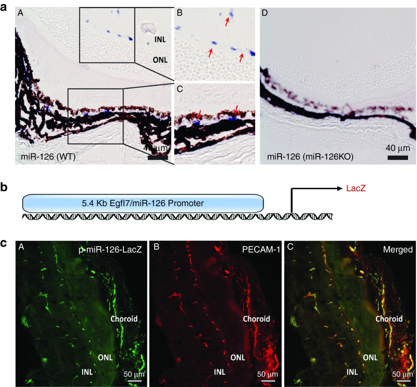
Figure 1.
EC-enriched miR-126 expression and miR-126/Egfl7 promoter activity in the mouse retina. (a) Section in situ detection of miR-126-3p expression in retinal and choroid vasculature in WT mice (A). (B) and (C) are the magnification of the boxed regions in (A). miR-126−/− retina (D) was used as negative control for specificity of the probe. INL: inner nuclear layer. ONL: outer nuclear layer. Scale bar equals to 40 µm. (b) Schematic pmiR-126/Egfl7-LacZ construct containing LacZ reporter driven by a 5.4kb miR-126/Egfl7 promoter. (c) LacZ and PECAM-1 co-staining of the retina cross sections in 4-week old pmiR-126/Egfl7-LacZ transgenic mice showing co-localized expression of LacZ and PECAM-1 in the retinal and choroidal vasculature. (A) LacZ antibody staining; (B) PECAM-1 staining; (C) Merged picture. Scale bar equals to 50 µm.

