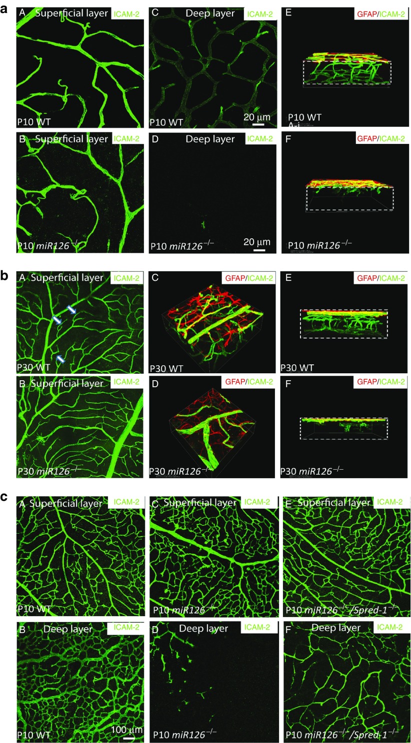
Figure 2.
Retinal vascular phenotype in miR-126−/− mice and its rescue by Spred-1 knockout. (a) Representative ICAM-2 (green) and glial fibrillary protein (GFAP) (red) staining of a postnatal day 10 (P10) WT and a miR-126−/− flatmount retina showing defective retinal vascular sprouting in miR-126−/− mice. A, C, and E are ICAM-2 staining of the retinal superficial layer, deep layer, and 3-D reconstruction of the retinas in WT mice; while B, D and E are the ICAM-2 staining of the corresponding regions in miR-126−/− mice. Boxes in E and F indicate the retina with different layers visualized. Scale bar equals 20 µm. (b) GFAP and ICAM-2 co-staining of the P30 WT and a miR-126−/− flatmount retina showing that retinal vascular sprouting is not recovered in P30 miR-126−/− mice. A and B show the ICAM-2 staining of the superficial layer retinas. Arrows indicate significantly more branches in the WT mice compared to the miR-126−/− mice. C to F are 3D reconstruction of the P30 retinas from different angles after ICAM-2/GFAP staining. (c) Partial rescue of retinal angiogenic phenotype in miR-126−/− mice by knockout of its target gene Spred-1. ICAM-2 staining of the superficial (A, C, E) and deep layer (B, D, F) of P10 retinas from different genotypes indicated was shown. Scale bar equals 100 µm.

