Figure 4.
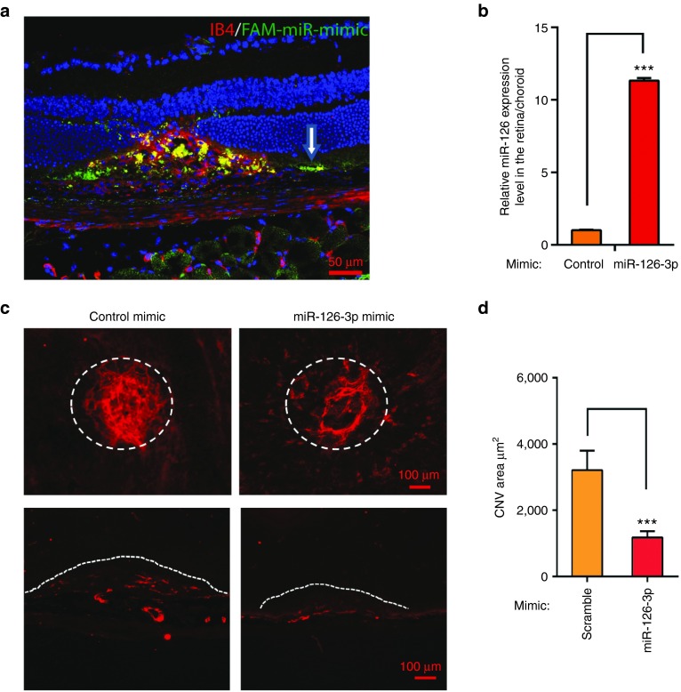
Repression of laser-induced CNV by miR-126-3p mimic in vivo. (a) Representative images of Isolectin-B4 staining at 3 days after laser injury subretinal injection of FAM labeled miRNA mimic. Note miRNA mimic taken up by RPE cells (white arrow) and choroidal vasculature (by IB4 and FAM co-staining) in the injured region. (b) Real-time PCR showing upregulation of miR-126-3p by miR-126-3p mimics in the posterior eyes. ***, P < 0.001. (c) Representative images after ICAM-2 staining showing repression of laser-induced CNV by miR-126-3p mimics compared to control mimics. Circled regions are the CNV areas. The bottom pictures show representative ICAM-2 staining of the injured regions after cross sectioning. miRNA mimic treatments were indicated and the lesion areas were labeled by dashed lines. Scale bar = 100 µm. (d) Quantification of CNV area (µm2) in c. ***, P < 0.001.

