Figure 6.
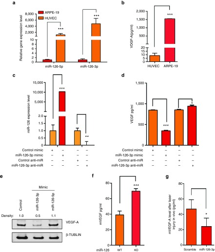
Regulation of VEGF expression by miR-126-3p in RPE cells. (a) Low expression of miR-126-3p and miR-126-5p in ARPE-19 cells compared to HUVECs as shown by qRT-PCR after normalized to U6. ***, P < .001. (b) Much higher secretion of VEGF-A by ARPE-19 cells compared to HUVEC cells as shown by ELISA analysis. ***, P < 0.001. (c) Upregulation and silencing of miR-126-3p in ARPE-19 cells by miR-126-3p mimic and LNA-modified antimiR, respectively. **, P < 0.01; ***, P < 0.001. (d) ELISA analyses showing regulation of VEGF-A by miR-126 mimic and anti-miR in ARPE-19 cells. *, P < 0.05; ***, P < 0.001. (e) Repression of VEGF-A by miR-126-3p but not miR-126-5p mimic in ARPE-19 cells as revealed by Western blot analysis. Density of the bands was noted. β-TUBULIN was used as control. (f) Elevated VEGF-A protein level as revealed by ELISA analysis in isolated miR-126−/− RPE cells compare to the WT RPE cells. (g) Repression of VEGF-A in the retina by miR-126-3p mimic injection after laser injury in vivo. *, P < 0.05.

