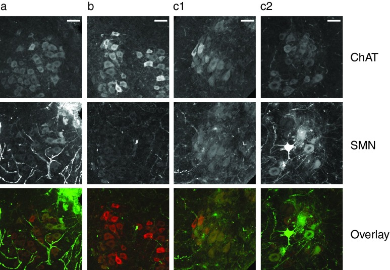
Figure 6.
Detection of SMN protein in spinal cord motoneurons of Spinal Muscular Atrophy (SMA) mice. Spinal cord sections from 10 day old mice were immunolabelled with anti-choline acetyl transferase (ChAT) to identify cholinergic neurons (top) and anti-SMN (middle) to reveal the level and distribution of SMN protein. The lowest row shows an overlay of the ChAT (red) and SMN (green) signals. (a) Smn1 +/− carrier mouse injected with phosphate-buffered saline. (b) Smn1 −/− disease mouse injected with PBS. (c1, c2) Smn1 −/− disease mice injected with scAAV9-4xsU7. Bar size = 50 µm.

