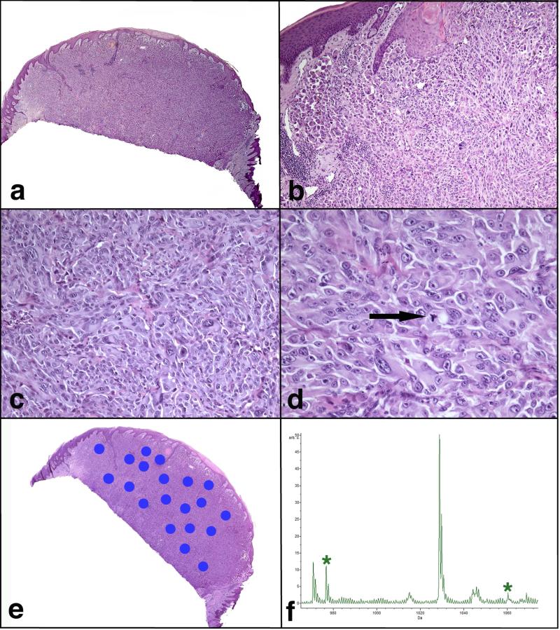
FIGURE 2.
Atypical Spitzoid Neoplasm diagnosed as Spitzoid melanoma by histopathological examination and as Spitz nevus by IMS. Patient #106, a 12-year-old boy with a nodule on the left lower leg, was rendered a histopathological diagnosis of SM with a tumor thickness of 3.7 mm. The lesion was ulcerated and there were 3 mitotic figures per mm2. This lesion was classified as SN by IMS. This patient (clinical Group 1c) had 1/1 SLN positive and no positive lymph nodes on completion lymphadenectomy. The patient was alive and free of disease 6 years after the diagnosis. He died in a car accident at the age of 18. a) Large nodular and poorly defined melanocytic lesion. b) There is mostly intradermal melanocytic proliferation with only focal junctional component. The epidermis is hyperplastic and shows hypergranulosis and hyperkeratosis. c) The proliferation is dense, forming sheets of melanocytes. d) The melanocytes are large, pleomorphic, with vesicular nuclei, prominent purple nucleoli and abundant eosinophilic cytoplasm. Mitotic figures are noted throughout the lesion (arrow). e) A scanned image of the specimen containing areas marked for Imaging Mass Spectrometry analysis. f) Average spectrum of the selected m/z range 965-1075. Two peaks at m/z 976.5 and m/z 1060.2, which are part of the classifier, are marked by *.

