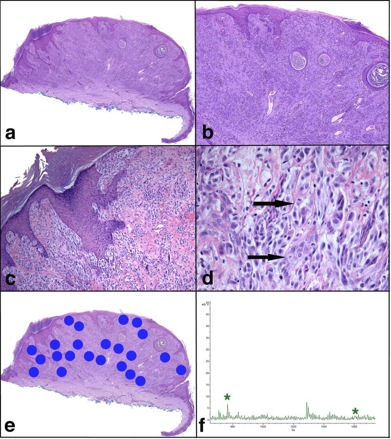
FIGURE 3.
Atypical Spitzoid Neoplasm with a diagnosis of ASN by histopathological examination and of Spitz nevus by IMS. Patient #97 is a 48-year-old woman with a lesion from the right volar forearm diagnosed histopathologically as ASN with tumor thickness of 2.25 mm and 1 mitosis per mm2. The lesion was classified as SN by IMS. This patient (clinical Group 1a) was alive and free of disease 5.5 years after the diagnosis. a) Broad and asymmetric proliferation of melanocytes. b) The epidermis is hyperplastic and shows hypergranulosis and hyperkeratosis. The majority of the lesion is intradermal and shows nests and fascicles of melanocytes. c) There are irregular nests of melanocytes in the epidermis, which vary in size and shape and are not equidistant from one another. Melanocytic nests are also seen down adnexal epithelium and in the dermis. d) The melanocytes are large, pleomorphic, some with hyperchromatic nuclei and abundant pale cytoplasm. Two mitotic figures are designated by arrows. e) A scanned image of the specimen containing areas marked for Imaging Mass Spectrometry analysis. f) Average spectrum of the selected m/z range 965-1075. Two peaks at m/z 976.5 and m/z 1060.2, which are part of the classifier, are marked by *.

