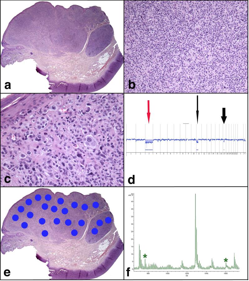
FIGURE 4.
Atypical Spitzoid Neoplasm diagnosed as Spitz nevus by both, histopathological examination and IMS. Patient #35 (clinical Group 2), a 58-year-old man, with a lesion from the ear diagnosed as favoring SN on histopathological assessment, was also diagnosed as SN by IMS. This lesion was completely excised. Fifteen years later there was a recurrence at the site of the primary lesion, which was also excised and diagnosed histopathologically as favoring a recurrent SN. Subsequent follow-up for another 10 years (25 years total) did not show any further recurrences or metastases. a) Large, asymmetric and somewhat multi-lobular melanocytic lesion. b) Very dense, sheet – like proliferation of melanocytes in the dermis. c) Single large melanocytes are seen at the dermal epidermal junction. In the underlying dermis there are large pleomorphic melanocytes with vesicular nuclei and abundant eosinophilic cytoplasm, focally forming small nests. d) An aCGH analysis shows loss of chromosome 3 (red arrow) and partial loss of chromosome 11p (long black arrow) and 17p (short black arrow), findings not entirely sufficient to support the diagnosis of melanoma. e) A scanned image of the specimen containing areas marked for Imaging Mass Spectrometry analysis. f) Average spectrum of the selected m/z range 965-1075. Two peaks at m/z 976.5 and m/z 1060.2, which are part of the classifier, are marked by *.

