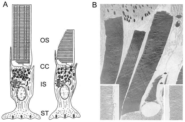
Figure 1. The OS in context.
A) Illustrations of rod (left) and cone (right) frog photoreceptors; adapted from (Bok, 1985). In each subtype, the light-sensitive OS is attached to the inner segment (IS) via the connecting cilium (CC). B) Transmission electron microscopy of adult frog OSs; three rod and one cone OSs are present. Apical processes (small arrows) and a phagosome containing shed disks (large arrow) are present. Insets show enlarged images of disk rims (left) and edges (right) from a cone OS. Reproduced with permission from (Kinney and Fisher, 1978a).

