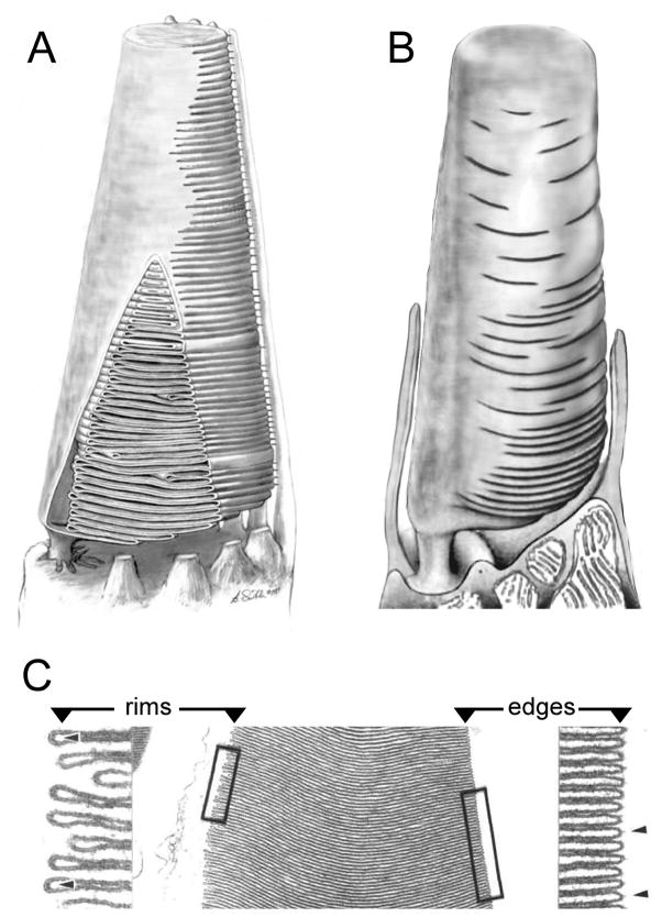
Figure 2. OS disk membrane topology varies by cell subtype, species, and disk age.
Renderings of cone photoreceptor OSs (A, B; not to scale). A) frog cone adapted from (Weber et al., 2011). Frog cone OSs possess a strong basal-to-distal taper. A portion of the plasma membrane has been cut away to reveal the internal structure. The plasma membrane opposite the eccentrically positioned cilium is pleated to form a large stack of disks that are further defined by the presence of rims, which partially bound the pleats and begin the process of defining a new compartment. The apertures that expose disk surfaces to the extracellular milieu extend about halfway around the disk boundaries, and are aligned along the length of the OS to those of adjacent disks. B) Mammalian cone adapted from (Anderson et al., 1978). Mammalian cone OSs possess more subtle basal-to-distal tapers than those present in frog cones. Like frog cone OSs, mammalian cone OSs also possess disks that remain in continuity with the plasma membrane. In this case however, the boundary of each mature disk is largely differentiated from the plasma membrane as a rim, so that the disks are largely internalized and only relatively small apertures remain open to the extracellular milieu. Moreover, these apertures are not aligned along the OS length, and make topological interpretation of sectioned tissue quite challenging. C) Transverse sections of frog cone OSs illustrate the distinction between U-shaped disk edges (right) and hairpin-shaped disk rims (left) that are present in the partially-internalized disks prevalent in cones. Adapted from (Corless and Fetter, 1987).

