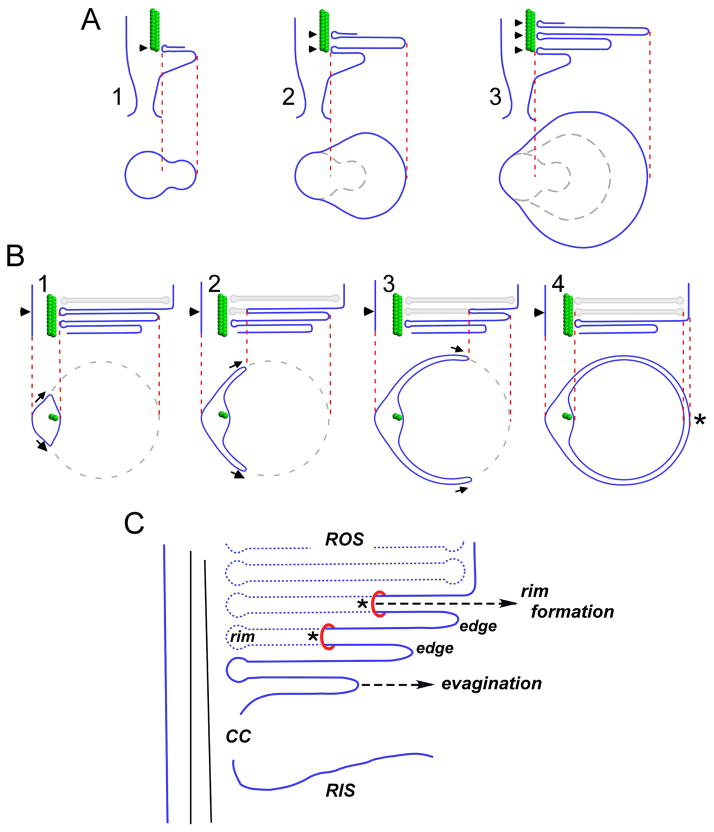
Figure 4. Steinberg et al. two-step (A, B) model for OS disk morphogenesis.
A) Step1 - growth of the ciliary plasma membrane creates an evagination with an upper surface anchored at an axonemal microtubule (A.1). Anchoring of the evagination lower surface allows for initiation of a new evagination (A.2). Evagination translation towards the cilium distal tip is accompanied by flattening and growth to mature disk diameter (A.3). At this stage, disks are bounded by edges; rims are absent, except for those regions immediately adjacent to the axonemal microtubules (arrowheads). Below: Tangential views sectioned through of each new evagination (arrowheads in longitudinal views). B) Step 2 - disk internalization begins by expansion of the rim region that anchors evaginations to the axoneme. Above: longitudinal views show expansion of the disk rim from left to right across one open disk (arrowheads). Below: tangential views sectioned through a single disk (arrowheads) undergoing internalization. The disk rim present at the axoneme advances bilaterally, in parallel with the enclosing plasma membrane at two growth points (arrows). Disk rim growth is proposed to occur by membrane addition. Completion of rim formation and disk internalization occurs opposite the axoneme (asterisk), and requires a membrane fission event to sever disk-plasma membrane continuity. Adapted from (Steinberg et al., 1980). C) A view of disk morphogenesis that emphasizes disk rim formation via membrane addition. Asterisks illustrate growth points, at which the addition of new plasma membrane allows rim formation to advance. Adapted from (Arikawa et al., 1992).

