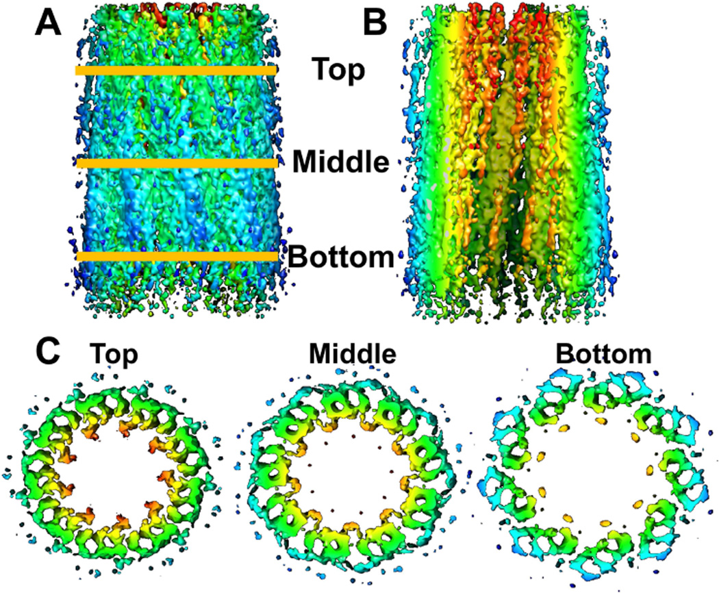
Figure 4.
Enhancement of cryo-ET reconstruction of a daughter centriole by sub-tomogram averaging. A. External view perpendicular to the central axis; B. cut-away view with outer half removed to reveal internal structures; C. Cross-sectional views of slices taken at the indicated axial positions. Colors indicate distance from central axis, orange (closest) to blue (furthest).

