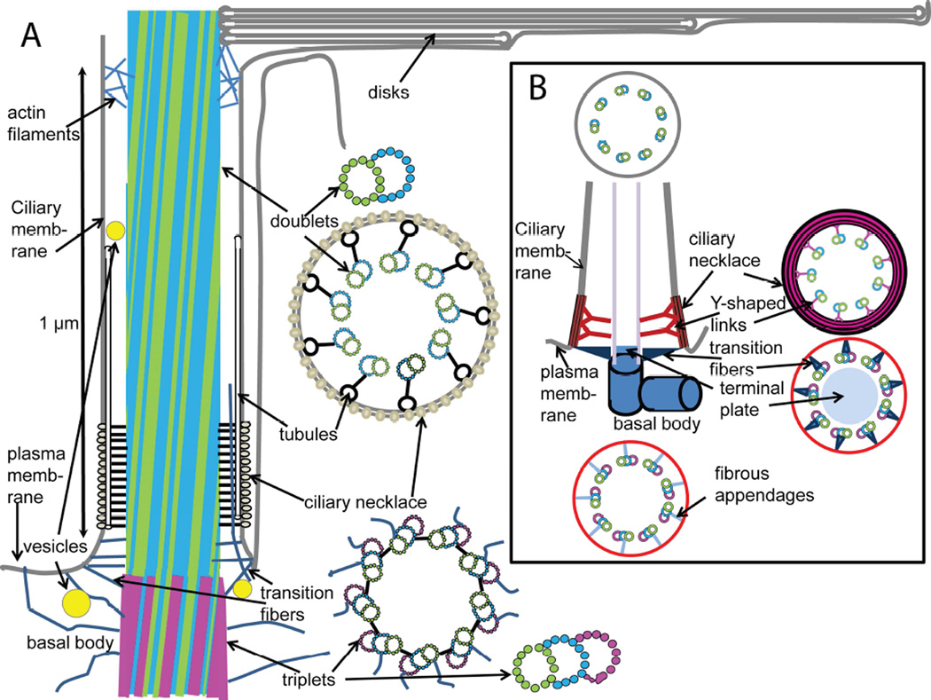
Figure 8.
Alternative models of basal body and connecting cilium structure based on cryo-electron tomography of isolated mouse rods. Panel A shows a model for the structural organization of the regions of the rod cell adjacent to the connecting cilium, based on available structural data, largely from various EM techniques. Distinctive features include: intersection of fibers emanating from the axoneme with tubules running along the length of the transition zone giving rise to the characteristic “Y-shaped links” in cross section; absence of clearly-defined triangular “alar sheet” transition fibers, and presence of multiple filaments connecting the microtubules to the plasma and ciliary membranes; vesicles adjacent to and within the lumen of the connecting cilium; filaments extending from the cytoplasm into the cilium lumen; thinning of basal body from minus (triplet) to plus (doublet) end; slight tilt of microtubules with respect to ciliary axis; envelopment of basal disks within plasma membrane (not necessarily to exclusion of some regions of continuity; see text). The arrangement of tubulin dimer units (circles) in doublet microtubules is based on the structure determined by cryo-ET and sub-tomogram averaging of axonemes from Chlamydomonas reinhardtii flagella (Nicastro et al., 2011), and that of the triplet microtubules is based on the basal body structure determined using similar approaches to basal bodies from the same organism (Li et al., 2012). The arrangement of the doublet and triplet microtubules is based on cryo-electron tomography and sub-tomogram averaging of mouse rod centrioles (Figs. 3 and 4 and Zhixian Zhang, Michael F. Schmid, Feng He, Theodore Wensel, unpublished observations). At the base of the outer segment, actin filaments are found within the lumen of the axoneme, and extending out between microtubules to contact the disk membranes (Chaitin and Bok, 1986; Chaitin and Burnside, 1989; Chaitin et al., 1984). Not shown is the daughter centriole invariably associated with the depicted structures. Panel B depicts a standard model of primary cilia, based on reference (Szymanska and Johnson, 2012).

