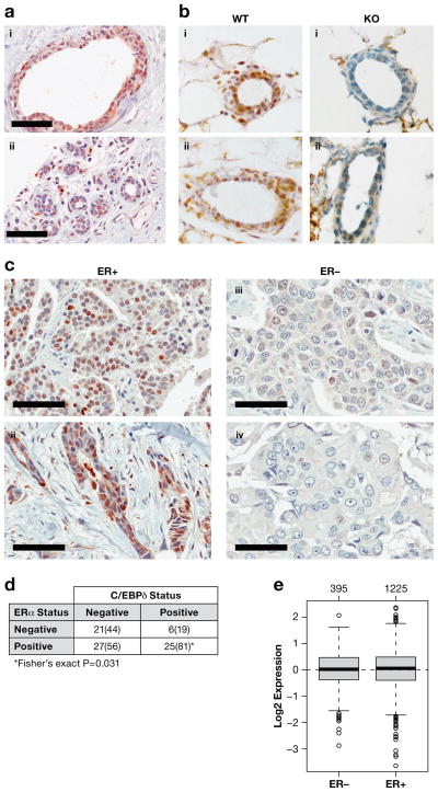
Figure 1. C/EBPδ is expressed in normal breast epithelial cells and ER+ breast cancer.
A) Immunohistochemistry of C/EBPδ of two independent human breast tissue specimen (scale bar = 60 μm). B) Immunohistochemistry of C/EBPδ on frozen sections of abdominal mammary glands from 4–month old Cebpd wild-type (WT) and null mutant (KO) mice at diestrous, two independent specimen each (i–ii). C) Immunohistochemistry of C/EBPδ in human breast cancer tissues with ER status as indicated. i: ductal papillary adenocarcinoma; ii–iv: invasive ductal carcinoma (scale bar = 60 μm). D) Correlation analysis of ERα and nuclear C/EBPδ staining in carcinoma cells of breast cancer tissue microarray 1 (TMA-1). Number of specimen (percentages for C/EBPδ in parenthesis) scored as positive or negative for C/EBPδ and ERα are shown. See Supplementary Figure S3A for distribution of C/EBPδ staining frequency and intensity. E) CEBPD mRNA expression in breast cancer tissues according to ER status as analyzed by GOBO (http://co.bmc.lu.se/gobo)43. The number of samples per group is shown above the plots.

