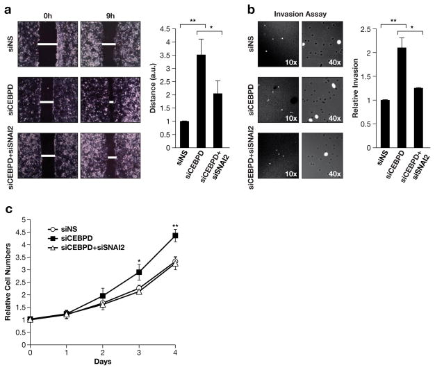
Figure 4. Silencing of SNAI2 expression reverts the phenotype of C/EBPδ-depleted MCF-7 cells.
A) Cell migration assay. MCF-7 cells were transfected with the indicated siRNAs and grown with culture inserts until confluent. Representative images (left) are shown at the indicated time after removal of the insert along with quantification (right) of wound closure from three independent experiments each done in triplicates (*P<0.05, ** P<0.01). B) Transwell invasion assay. MCF-7 cells treated as in panel A were plated without serum in Matrigel invasion chambers. Cells that migrated to the side with serum-containing medium after 36 h were stained with DAPI and counted (n=3,* P<0.05, ** P<0.01). C) Analysis of cell population growth/viability by vital dye staining (Alamar blue) of MCF-7 cells with transient knockdown of C/EBPδ (siCEBPD), SNAI2 (siSNAI2), or control (siNS) (n=3, ** P<0.05). ** P<0.01).

