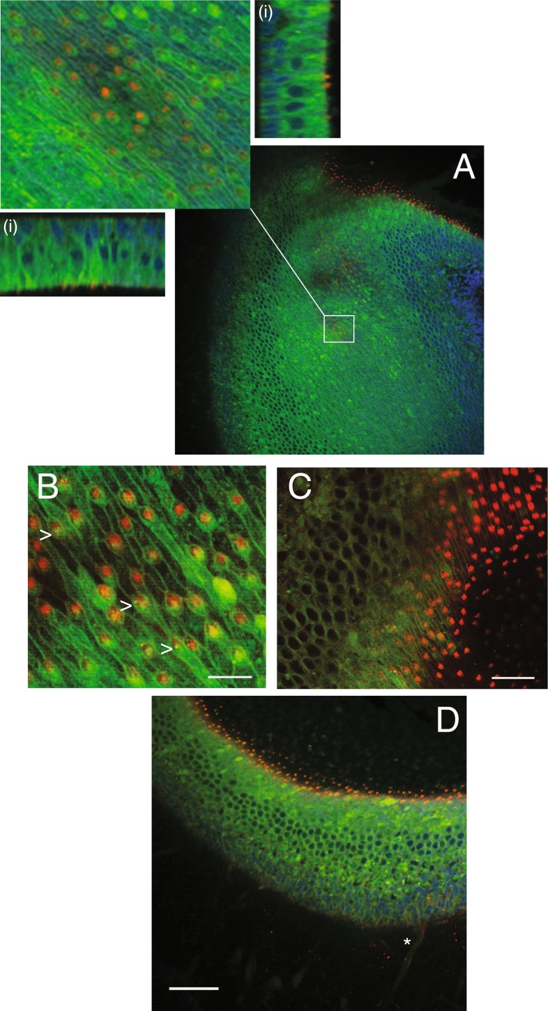
FIG. 3.
Distribution of BX-primed GAGs in the utricle and saccule. Utricles were stained with DAPI (blue) and phalloidin (red) to visualize nuclei and stereocilia, respectively. A Two-photon projection of a z-stack image demonstrates the presence of BX-GAGs (green) in several structures in the utricular epithelium. Orthographic projections of an enlarged region of the utricular epithelium demonstrating the extent of BX-GAGs throughout utricular hair cells and supporting cells (i). B BX-GAGs at the apical face of hair cells formed a meshwork glycocalyx between cells that aligned with the local polarity of hair bundles. A fluorescent BX-GAG glycocalyx developed around each kinocilia (arrows; scale bar 10 μm). Stereocilia bundles (labeled red) project away from the cell bodies in the same orientation as the kinocilia resulting in the yellow appearance of green glycocalyx enveloped kinocilia (arrow). C Alternative lower magnification cross-sectional view of B, showing stereocilia bundles (red) projecting from the hair cells (green; scale bar 20 μm). D Cross-sectional view of the hair cells, supporting cells, kinocilia, and an afferent nerve (star) in a representative saccule (scale bar 50 μm)

