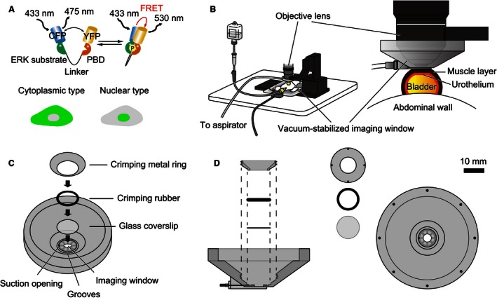
Figure 1.

Experimental set‐up for intravital imaging of the mouse bladder. (A) The mode of action of a Förster/fluorescence resonance energy transfer (FRET) biosensor for extracellular signal‐regulated kinase (ERK) activity, EKAREV. Phosphorylation of the ERK substrate region within the biosensor induces conformational change, which brings yellow fluorescent protein (YFP) into close proximity of cyan fluorescent protein (CFP) and YFP, and thereby increases the level of FRET. The phosphate binding domain binds to the ERK substrate region in a phosphorylation‐dependent manner. Two types of biosensors, the EKAREV‐nuclear export signal and EKAREV‐nuclear localization signal, are in the cytoplasm and the nucleus, respectively. (B) Layout of the intravital imaging system for the mouse bladder. An anesthetized mouse is placed on an electric heat pad. The bladder is attached to the vacuum‐stabilized imaging window. (C) Detailed schemes of the vacuum‐stabilized imaging window. The bowl of the imaging window, which has a glass coverslip at the bottom, is designed to be filled with water for the water‐immersion objective. The suction opening is placed on the bottom surface of the imaging window to enable the aspirator to continuously suck on the bladder. (D) The side and top views of the vacuum‐stabilized imaging window. The crimping metal ring and a crimping rubber are used to seal the water reservoir. The crimping metal ring has four small pits to fit a special screwdriver.
