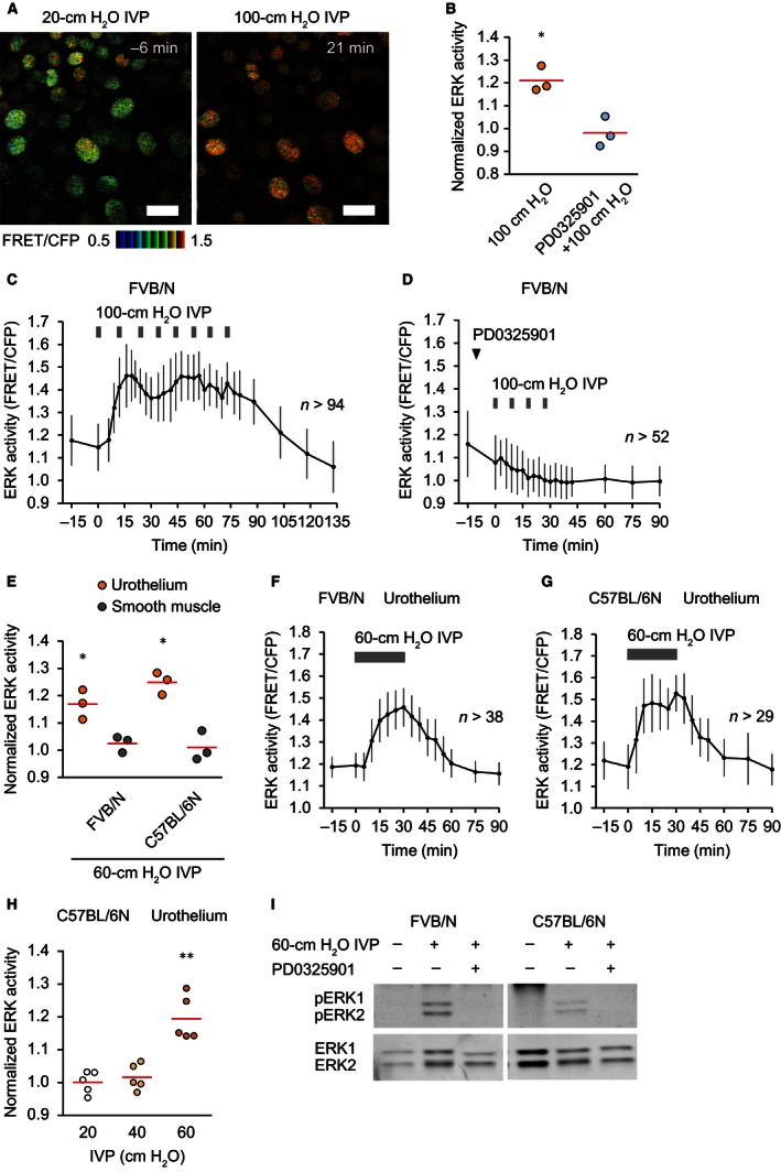
Figure 3.

Extracellular signal‐regulated kinase (ERK) activation by bladder distention with increased intravesical pressure (IVP) in the urothelium. (A) Representative ratio images of high IVP‐induced ERK activation in the urothelium of an Eisuke‐NLS‐FVB mouse (Video S3). Scale bar = 20 μm. (B) Summary of the relative change in urothelial ERK activity of Eisuke‐NLS‐FVB mice by intermittent 1‐min 100‐cm H2O IVP and the effect of an MEK inhibitor, PD0325901 (5 mg kg−1), on high IVP‐induced ERK activation (n = 3 for each). ERK activity was measured at baseline (0 min) and 15 min after application of the first 1‐min of 100‐cm H2O IVP. PD0325901 was injected intravenously 10 min before application of high IVP. Circles and red lines indicate normalized ERK activity in each experiment and the mean values, respectively. (C and D) Representative graphs of the change in urothelial ERK activity by intermittent 1‐min 100‐cm H2O IVP (C) and the effect of PD0325901 on high IVP‐induced ERK activation (D). n, number of cells analyzed at each time point. (E) Summary of the relative change in ERK activity by 30‐min 60‐cm H2O IVP in the urothelium and the smooth muscle of both Eisuke‐NLS‐FVB and Eisuke‐NLS mice (n = 3 for each). ERK activity was measured at baseline (0 min) and 30 min after application of high IVP. Circles and red lines indicate normalized ERK activity in each experiment and the mean values, respectively. (F and G) Representative graphs of the change in urothelial ERK activity of Eisuke‐NLS‐FVB (F) and Eisuke‐NLS mice (G) by 30‐min 60‐cm H2O IVP. n, number of cells analyzed at each time point. (H) Summary of the relative change in urothelial ERK activity of Eisuke‐NLS mice by 30‐min 20‐, 40‐, or 60‐cm H2O IVP (n = 5 for each). ERK activity was measured at baseline (0 min) and 30 min after application of IVP. Circles and red lines indicate normalized ERK activity in each experiment and the mean values, respectively. (I) Western blot analysis showing ERK1/2 and phospho‐ERK1/2 in the isolated urothelium of Eisuke‐NLS‐FVB and Eisuke‐NLS mice. Bladders were extracted without application of high IVP (left lane), after application of 30‐min 60‐cm H2O IVP (middle lane) and after application of 30‐min 60‐cm H2O IVP with intravenous injection of PD0325901 (5 mg kg−1) 10 min before application of high IVP (right lane). *P < 0.05, **P < 0.01 compared to the control by a paired Student's t‐test.
