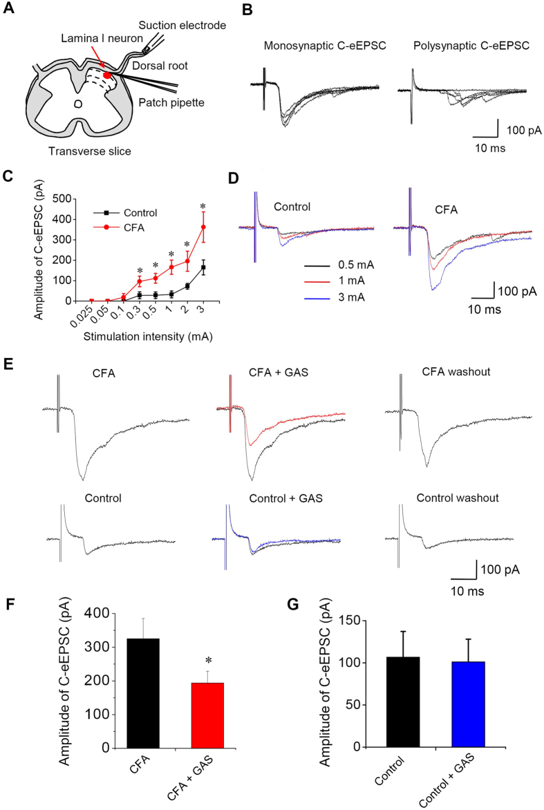
Figure 4. GAS preferentially inhibited spinal synaptic potentiation under inflammatory pain states but with intact basal synaptic transmission.
(A) Schematic diagram showing whole-cell patch-clamp recording from lamina I neurons in a spinal cord slice attached with a dorsal root. (B) Typical examples of monosynaptic C-eEPSCs and polysynaptic C-eEPSCs recorded in spinal lamina I neurons. (C) Input-output curves for synaptic transmission between C-fibers and spinal lamina I neurons in control and CFA-inflamed mice (n = 8, P < 0.05). (D) Typical traces of C-eEPSCs in response to increasing intensity of dorsal root stimulation from control and CFA-inflamed spinal slice. (E) Representative traces showing that bath application of GAS (300 μM) reversibly inhibited the peak amplitude of C-eEPSCs recorded from CFA-inflamed spinal slice (upper panels) but not that from control spinal slice (lower panels). Quantitative summary of the effect of GAS on C-eEPSC in CFA-inflamed and control mice are plotted in (F) (n = 10, P < 0.05) and (G) (n = 7, P > 0.05), respectively. All data are represented as mean ± S.E.M. *P < 0.05.

