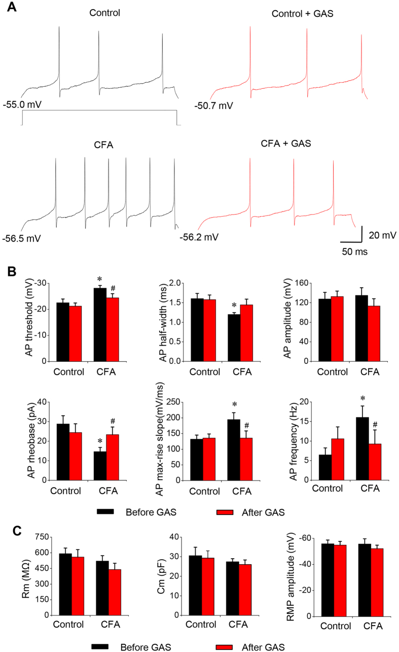
Figure 7. The inhibitory effect of GAS on the membrane properties and hyperexcitability of spinal lamina I neurons from CFA-inflamed mice.
(A) Representative traces showing the spike firings to a depolarizing current step prior to (left panels) and during GAS application (right panels) in spinal lamina I neurons from control (upper panels) and CFA-inflamed (lower panels) mice. Note that the traces before and after GAS application in each group were from the same neuron. (B) Quantitative analysis showing the active membrane properties, such as AP threshold, amplitude, half-width, rheobase, max-rise slope and firing frequency in spinal lamina I neurons from control and CFA-inflamed mice prior to (black column) and during GAS application (red column). Note that lamina I neurons from CFA-inflamed mice displayed lowered AP thresholds, shortened AP half-widths, reduced rheobase, increased AP rise slope and increased firing frequency compared to controls. These changes in CFA-inflamed mice were normalized by GAS. In contrast, GAS exerted no effect on the membrane properties of lamina I neurons from control mice. (C) Passive membrane properties of lamina I neurons from control and CFA-inflamed mice as well as the action of GAS on these passive membrane properties. All data are represented as mean ± S.E.M. *P < 0.05 compared to the control mice. #P < 0.05 compared to prior to GAS application in CFA-inflamed mice.

