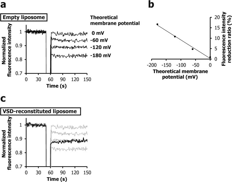
Figure 1. Measurement of membrane potentials of liposomes.
(a) Voltage dependence of the fluorescence intensity of empty liposomes. (b) Relationship between the theoretical membrane potential formed on empty liposomes and the fluorescence intensity reduction ratio of di-4-ANEPPS (panel (a)). (c) Fluorescence time-course for the liposomes reconstituted with a VSD mutant, V42C/I131C, where the K+ concentration ratio was adjusted to form a −187 mV theoretical membrane potential (black line). The results of (a) are shown by gray lines. The potentials corresponded to −140 and −124 mV at a few and 90 s after the addition of valinomycin, respectively.

