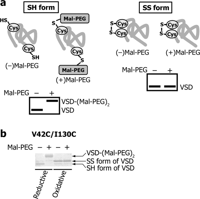
Figure 3. Detection of an intramolecular SS-bond.
(a) Schematic diagram of the intramolecular SS-bond detection. The SH form of the VSD mutant is PEGylated and exhibited a significant mobility shift (left), while the SS form of the VSD mutant is not PEGylated, leaving the mobility unchanged (right). (b) SDS-PAGE mobility of the VSD double Cys mutant (V42C/I130C) under reductive or oxidative conditions in the presence or absence of mal-PEG.

