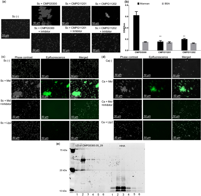
Figure 3. Agglutination assay of L. plantarum CMPG5300, its mutant derivatives and the lectin domain of Cmpg5300.05_29 with S. cerevisiae and C. albicans.
(a) Phase contrast images of the agglutination assay of S. cerevisiae BY4741 (Sc) incubated with L. plantarum CMPG5300, CMPG11201 mutant and CMPG11202 mutant alone and in the presence of methyl-α-D-mannopyranoside inhibitor. For each strain and condition tested, the experiment was performed at least three times. The images shown are representative of at least five imaging fields. (b) Binding of biotin-labeled cells of the wild-type strain L. plantarum CMPG5300, mutant CMPG11201 and complemented strain CMPG11202 to yeast mannan. The OD at 405 nm reflects the binding efficiency of the bacterial strains. BSA was used as a negative control. The dataset comparisons (mutant pairwise to wild-type) are considered significant (p < 0.01 indicated with two asterisks in the figure). The dataset comparison (complemented mutant pairwise to the mutant strain) show no significant differences. (c) Fluorescent images of the agglutination assay of S. cerevisiae BY4741 (Sc) and (d) C. albicans SC5314 (Ca) in the presence of the FITC labeled lectin domain of Cmpg5300.05_29, FITC labeled lectin domain of Cmpg5300.05_29 in the presence of methyl-α-D-mannopyranoside and lectin-like protein 2 (Llp2) from L. rhamnosus GG used as control. For each strain and condition tested, the experiment was performed at least three times. The images shown are representative of at least five imaging fields. (e) Proteins that bound to uncoated sepharose beads (lane 1, used as negative control), sepharose beads coated with various sugars: mannan (lane 2), mannose (lane 3), glucose (lane 4), fucose (lane 5), GlcNAc (lane 6) as separated by SDS-PAGE. The first six lanes (LD of CMPG5300.05_29) represent the lectin domain of CMPG5300.05_29 and the next six lanes represent HHA (used as control).

