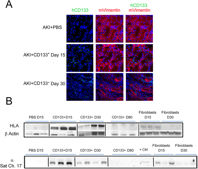
Figure 5. Intravenously injected CD133+ cells localization in kidneys of AKI SCID mice.
(A) Representative confocal micrographs showing the presence of CD133+ cells localized in the interstitium and tubules within the kidney of AKI mice at day 15 and day 30 after damage as evaluated by human CD133 (Green) and mouse Vimentin (red). Nuclei were counter-stained with DAPI (blue). Original magnification 400×. (B) HLA protein and whole genome DNA analysis (α-Sat ch17) of mice kidneys at day 15, day 30 and day 60 after glycerol injection. CD133+ cells were present up to day 30 as shown by immunofluorescence or protein/DNA analysis, whereas dermal fibroblasts were only detected at day 15. + Ctrl: positive control using human CD133+ cells. Lanes run on different gels are separated by a dark line.

