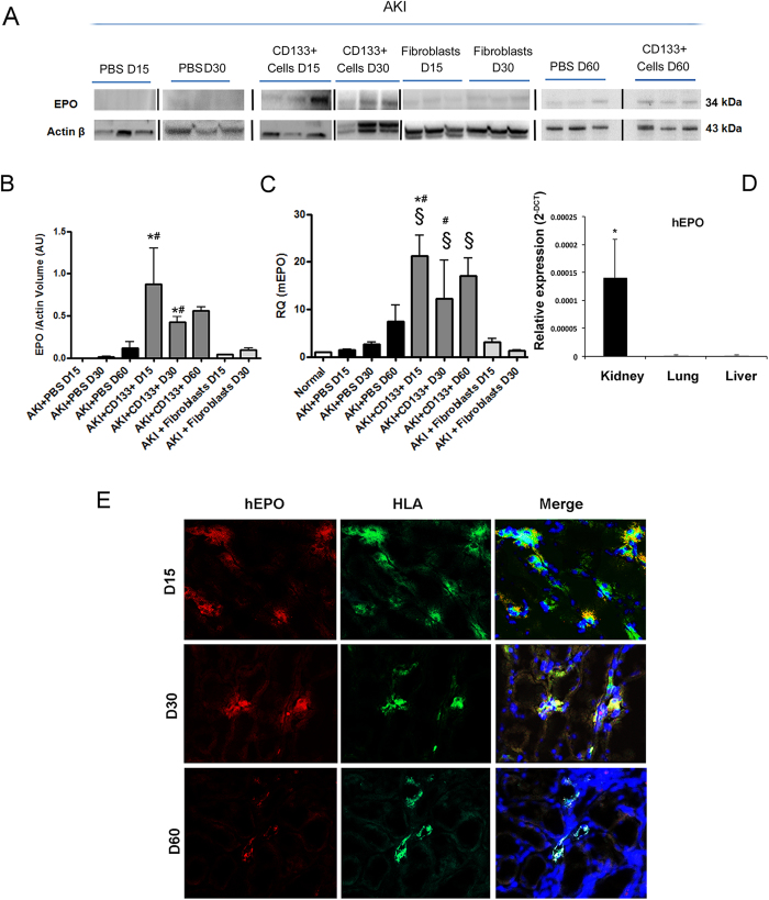
Figure 8. Effect of CD133+ cells on human and murine EPO.
(A,B) Western blot analysis and quantification of EPO protein in whole kidney lysate of mice treated with PBS, CD133+ cells or dermal fibroblasts at day 15, 30 and 60 after injury. Data are mean ± SD of three experiments analysing at least three different mice per group. Lanes run on different gels are separated by a dark line. (C) Quantitative RT-PCR analysis of mice EPO in whole kidney lysate of normal and AKI mice. Increased EPO levels are observed in CD133+ cell treated mice as compared to controls or fibroblasts. Data are normalized to GAPDH mRNA and to control and are expressed as mean ± SD of three to six different mice per group. Newman Keuls’ multicomparison test was performed: §p < 0.01 versus Normal; *p < 0.01 versus PBS, #p < 0.01 versus fibroblasts. (D) Quantitative RT-PCR analysis of human EPO in whole lysate of kidneys, lungs and livers of CD133-treated AKI mice at day 15. EPO levels were undetectable in lungs and liver. Data are reported as the relative expression (2−ΔCT). GAPDH was used as housekeeping gene. N = three mice. (E) Representative confocal micrographs showing the presence of human EPO (red) and HLA positive cells (green) within the kidney of AKI mice treated with CD133+ at day 15, 30 and day 60 after damage. Nuclei were counter-stained with DAPI (blue). Original magnification 400×.

