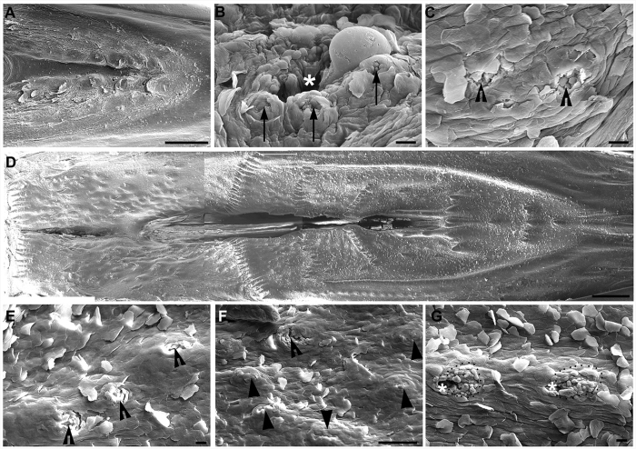
Figure 5. Scanning electron photomicrographs from the base of the oral cavity and palate in a P3 male chicken.
(A) Low magnification of the base of the oral cavity showing the structure and topography of the oral surface. (B) A taste bud cluster showing 3 taste buds (arrows) located around a salivary gland opening (asterisks). (C) Representative images of taste buds (open arrowheads) that were not close to a salivary gland opening. (D) Low magnification image of the palate showing the topography of the oral surface. (E,F) Taste buds (open arrowheads) located in the absence of a salivary gland opening in the palate. Solid arrowheads in F point to tissue protrusions that were probably developing taste buds without an obvious taste pore. (G) Taste buds (dotted outlines) surrounding the salivary gland opening (asterisks) in the palate. Scale bars: 1 mm for (A) and (D); 20 μm for (B,C), (E,G); 100 μm for (F).

