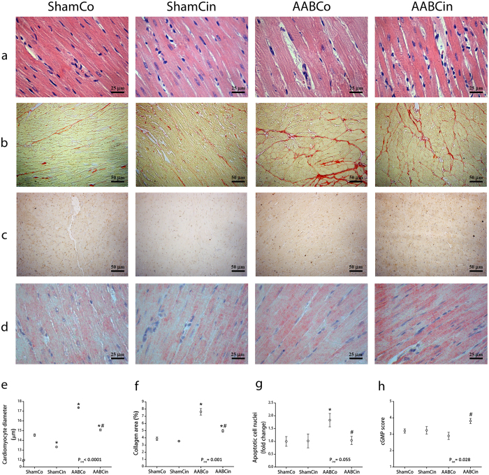
Figure 3. Histological alterations in pressure overload are blunted by Cinaciguat.
Differences between the groups are illustrated in representative photomicrographs of left ventricular (LV) myocardial sections with haematoxylin-eosin (a) and Picrosirius red staining (b), TUNEL (c) and cGMP immunohistochemistry (d). Average cardiomyocyte diameter (e) and collagen area of subendocardial LV myocardium (f) was significantly elevated in the AABCo group compared to ShamCo, both of which alterations were significantly decreased following Cinaciguat treatment. TUNEL staining revealed a significant increase in the number of apoptotic cell nuclei in the AABCo group compared to ShamCo and AABCin (g). cGMP score (h) was significantly higher in the AABCin group than in the AABCo animals. pint: interaction p value *p < 0.05 vs. ShamCo; #p < 0.05 vs. AABCo.

