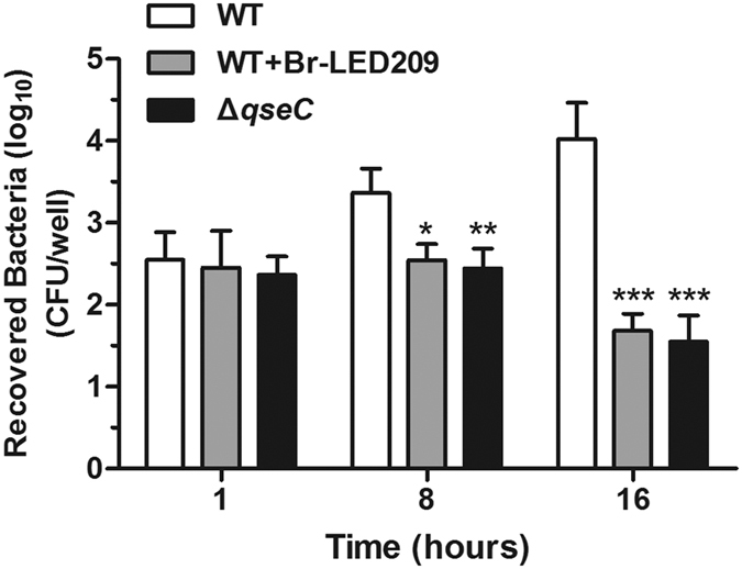
Figure 6. Elimination of intracellular bacteria from macrophages was enhanced by blocking QseC.

S. Typhimurium were cultured at 37 °C with shaking overnight and opsonized in DMEM and 10% normal mouse serum for 20 min. Macrophages were infected with opsonized WT S. Typhimurium or the qseC mutant at MOI of 25:1 for 30 min. The cells were washed with PBS and treated with 100 μg/ml gentamicin for 1 h to kill extracellular bacteria. Then, cell culture medium was replaced with 10 μg/ml gentamicin for the remainder of the experiment. To count the number of intracellular bacteria, macrophages were lysed with 1% Triton X-100. Bacteria were diluted and plated on LB medium plates to calculate CFU at the indicated time points. (*P < 0.05, **P < 0.01, ***P < 0.001 vs. WT in two-way ANOVA, n = 3).
