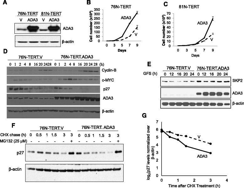
Fig. 2.

Exogenous overexpression of ADA3 leads to increased proliferation. a Western blotting of cells overexpressing ADA3. V = vector. 76 N-TERT cell line (b) or 81 N-TERT cell line (c) expressing an empty vector (V) or ADA3 were plated at a density of 1 × 10⁴ and then counted using a hemocytometer on alternate days to measure proliferation. d Western blotting of cell lysates from synchronized (0 time point) or cells released from synchrony (1-28 hours) were immunoblotted with indicated antibodies. β-actin was used as a loading control. e Western blotting of cell lysates from synchronized (0 time point and indicated time points released from synchrony (1-24 hours) were immunoblotted with SKP2, ADA3 or β-actin (used as a loading control). f p27 half-life analysis. 76 N-TERT cells expressing vector or overexpressing ADA3 were treated with cycloheximide, and then cells lysates at indicated time points were immunoblotted with anti-p27 antibody. Last lane, 3 hour time point of cells were treated with MG132. g The intensity of p27 bands was quantified by densitometry, normalized to β-actin using ImageJ software, and then plotted against the time of cycloheximide treatment
