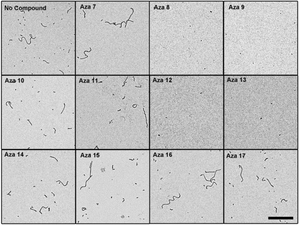
Figure 5. Electron microscopy of tau filament disassembly in the presence of azaphilone derivatives.
Tau polymerization reactions were performed with 2 µM tau and 75 µM arachidonic acid at room temperature. After 6 hours, 200 µM compound or equal volume of DMSO was added to the reactions. Aliquots of the reactions were prepared for negative stain electron microscopy. Representative images are shown for A) no compound control, B) aza-7, C) aza-8, D) aza-9, E) aza-10, F) aza-11, G) aza-12, H) aza-13, I) aza-14, J) aza-15, K) aza-16, and L) aza-17. The scale bar in the lower right panel represents 1 µm and is applicable for all images.

