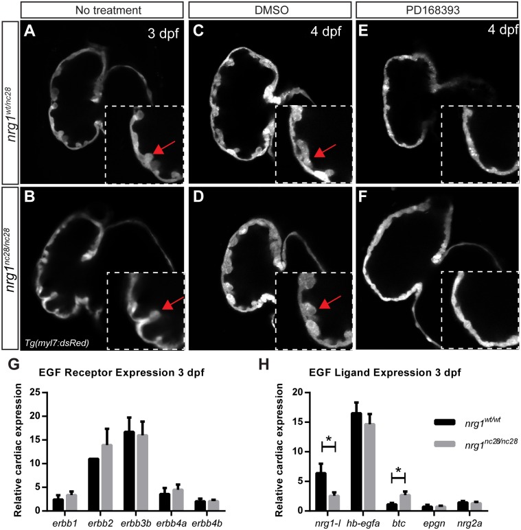
Fig 3. nrg1 mutants require ErbB2 tyrosine kinase activity to form trabeculae.
(A-F) Representative confocal optical mid-chamber slice of the ventricle at 3–4 dpf in larvae carrying Tg(myl7:dsRed) cardiomyocyte reporters. Boxes include high-resolution image of the outer curvature. Larvae were examined at (A-B) 3 dpf or (C-F) 4 dpf after treatment with (C-D) 1% DMSO or (E-F) 3.75 μM PD168393 from 2–4 dpf. Larvae were genotyped after imaging. Red arrows point to representative trabeculae. N ≥ 4 larvae for each condition and genotype. Relative expression levels of (G) EGF family receptor genes or (H) EGF family receptor ligand genes from isolated hearts of nrg1WT/WT and nrg1nc28/nc28 larvae at 3 dpf. N = 3–5 biological replicates with 30–60 hearts pooled per condition normalized to efl1a. N = 1 biological replicates with 30–60 hearts pooled for erbb2 normalized to efl1a. Student’s T-test mutant compared to wild type. Error bars are SEM. N≥3 biological replicates. *p≤0.05–0.01.

