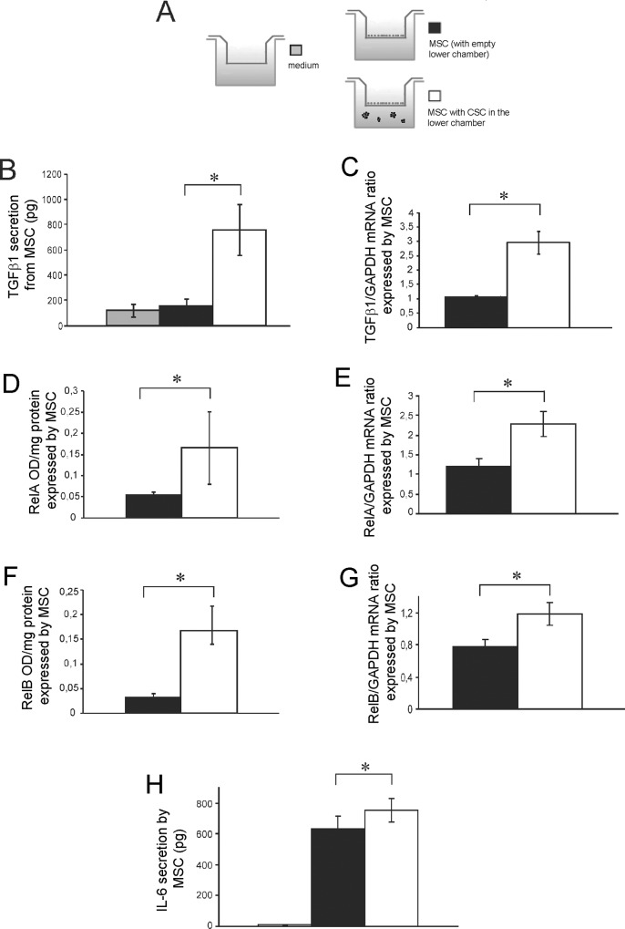
Fig 6. The presence of HOS-CSC enhance the secretion of TGFβ1 from MSC that, in turn. autocrinally induce the activation of inflammatory pathways.
(A) Scheme of the experiments used to evaluate the secreted proteins showed in the following panels. The secreted protein (only the supernatant in the MSC upper compartment was) (B) and mRNA expression (C) of TGFβ1 in MSC when co-cultured with HOS-CSC, after 3 days (*p<0.05). The presence of HOS-CSC, increased the expression levels in MSC of two genes of the NF-kB pathway, RelA (D for protein and E for mRNA) and RelB (F for protein and G for mRNA), as shown by direct NF-kB nuclear translocation (D and F panels) or transcriptional activation (E and G panels), *p<0.05. (H) When activated by the presence of HOS-CSC, MSC secreted higher amounts of IL-6, as assessed by ELISA assay, (*p<0.05).

