
Figure 2.
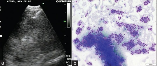
(a) Endobronchial ultrasound image of needle aspiration of the thyroid mass. (b) Cytopathological examination of the endobronchial ultrasound-guided transbronchial needle aspiration aspirate. May-Grünwald-Giemsa stain shows numerous microfollicles and few cohesive fragments of follicular cells. Original magnification × 100