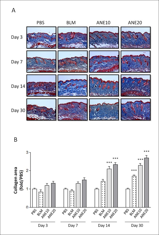
Fig 2. Histopathologic evaluation of the SC-injected ANE-induced changes in dermal collagen synthesis.
(A).The skin injection sites were examined on days 3, 7, 14, and 30by Masson’s trichrome staining. The sections were viewed under a microscope at 100x magnification. Scale bars indicate 100 μm. (B). the intensity of the blue color representing the collagen density was measured using IMAGE-Pro software. Each treatment group included at least 6 mice. The results are shown as the mean± SD. The levels of statistical significance are as follows: *P<0.05, **P<0.01 and ***P<0.001 relative to the control group.

