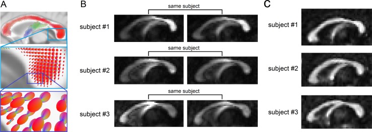
Fig 1. The uniqueness of local connectome structure revealed by the density of diffusing water.
(A) The spin distribution function (SDF) calculated from diffusion MRI quantifies the density of diffusing water along axonal fiber bundles. The magnitudes of the SDF at axonal directions provide density-based measurements to characterize axonal fiber bundles. (B) The density measurements obtained from the SDFs show individuality between-subjects #1, #2, and #3 (intensity scaled between [0 0.8]). The density of diffusing water varies substantially across different portions of the corpus callosum. The repeat measurements after 238 (subject #1), 191 (subject #2), and 198 (subject #3) days present a consistent pattern that captures individual variability. (C) In contrast to the SDF shown in (B), the fractional anisotropy derived from diffusivity shows no obvious individuality between the same subjects #1, #2, and #3 (intensity also scaled between [0 0.8]). This is due to the fact that diffusivity, which quantifies how fast water diffuses, does not vary a lot in normal axonal bundles.

