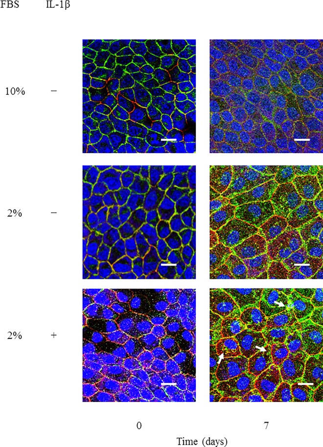
Fig 1. IL-1β induced a disruption in the localization of E-cadherin and ZO-1 and barrier function in MDCK cells.
Cells were treated with culture medium containing 10% or 2% FBS in the presence or absence of 50 pM IL-1β for the indicated time periods. Then, the cells were labeled with fluorescent dye for nuclear staining (blue, nuclei) and antibodies against E-cadherin (green) and ZO-1 (red). The IL-1β-treated cells displayed a loss of membrane localization of E-cadherin and ZO-1. Spotty cytoplasmic localization patterns of E-cadherin and ZO-1 (arrows) were observed in the IL-1β-treated cells.

