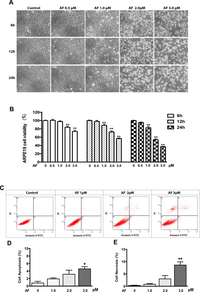
Fig 1. Auranofin inhibits ARPE-19 cells survival.
(A) Phase-contrast photomicrographs of ARPE-19 cells after 0–3.0 μM AF treatment for 6, 12 and 24 hours. Scale bar = 50μm. (B) ARPE-19 cells were treated with AF at 0–3.0 μM for 6, 12 and 24 hours. Cells viability was measured by an MTT assay. The cell viability is expressed as a percentage of control and data are from three independent experiments, n = 4. (C) ARPE-19 cells were treated with AF at 1.0 μM, 2.0 μM and 3.0 μM for 12 hours and stained with PI and Annexin V-FITC. Cells death was detected by flow cytometery (in dot plots, necrosis is only PI+ in area 1(A1), late apoptosis is annexin V++PI+ in area 2(A2), normal living is annexin V−+PI− in area 3 (A3), early apoptosis is annexin V+ in area 4 (A4)). (D) Quantification of ARPE-19 cell apoptosis (early apoptosis: annexin V+, late apoptosis: annexin V++PI+) induced by AF, n = 3. (E) Quantification of ARPE-19 cell necrosis (PI+) induced by AF, n = 3. All data are means ± SEM, *P<0.05, **P<0.01, compared with control.

