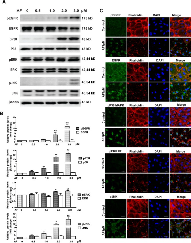
Fig 3. Auranofin induces phosphorylation of EGFR/MAPK.
(A) ARPE-19 cells were treated with different doses of AF for 12 hours. Cell lysates were subjected to Western blot for total and phosphorylated EGFR, P38MAPK, ERK, JNK, with respective antibodies. β-actin was used as an internal control. (B) Quantitative data of Western blot presented in panel A from three independent experiments. The levels of phosphorylated protein were compared with the control levels, *P < 0.05,**P < 0.01; the levels of total protein were compared with control levels,※P < 0.05,※※P < 0.01. (C) Immunofluorescence microphotographs of total and phosphorylated EGFR (green), phosphorylated P38MAPK (green), phosphorylated ERK(green), phosphorylated JNK(green) with phalloidin (red) and DAPI (blue) after ARPE-19 cells were treated with 2.0 μM AF for 12 hours. Scale bar = 20μm.

