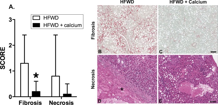
Fig 2. Histological scoring of fibrosis and necrosis.
A. Fibrosis as indicated by Sirius red-staining for collagen was significantly lower in the HFWD-Ca group (asterisk, Mann-Whitney U, p = 0.0007). Necrosis assessed in H&E stained sections was also lower in the HFWD-Ca group, although this difference was not significant. B-E. Representative histological features. Fibrosis (B and C) varied from increased pericellular (“chicken wire” pattern) to pericentral (lobular) deposition in Sirius red stained sections. Necrosis (D) consisted of areas of devitalization (asterisk) that most commonly occurred within large regenerative hyperplastic nodules (RH) in HFWD mice. (E) depicts liver from a HFWD-Ca mouse with steatosis but no necrosis for comparison. Hematoxylin and eosin staining. Scale bar = 100μm.

